Mouse serotonin neurons sort into five groups by brain function
A Cell study gives the first whole-brain map of serotonin projections in a vertebrate. Each of five neuron groups targets a set of functionally related regions.
Why it matters · Serotonin affects mood, movement and anxiety, and it is a major drug target. Its wiring was poorly understood until now. This map gives researchers a structure for testing what each branch of the system actually does.
Serotonin neurons in the mouse brain fall into five distinct groups, and each group sends its fibers to a set of functionally related brain regions. That is the main finding of a study published last month in Cell. It is the first projectome, or whole-brain map of connections, for the serotonin system of any vertebrate. Another team mapped the full serotonin system of a roundworm in 2023.
How the map was made
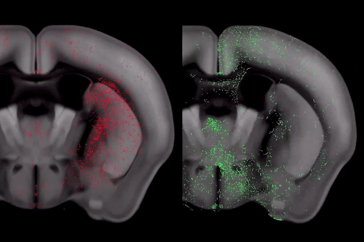
The team, which includes Stanford neurobiologist Liqun Luo, built on mouse work from 2018. They used viral-genetic tracing to follow serotonin neurons that start in the dorsal and median raphe, two clusters in the brainstem. Mice were injected with a virus that makes connected serotonin neurons light up across the whole brain. The researchers then imaged the brains and repeated the process until they had covered every area that serotonin reaches.
The five projection groups are:
- the hippocampal-entorhinal network
- the basal ganglia
- the cortex
- the medial interbrain (medial thalamus and hypothalamus)
- the brainstem and lateral thalamic nuclei
The authors argue that the system is organized by what its target regions do, not by how close they are. There are exceptions. The central and basolateral amygdala sit next to each other and both help regulate fear and emotion, yet they belong to different groups. That split matches their different developmental origins and cell types.