Roots Recoil, Implants Talk, Stars Stay Put · Morning Gist
Sunday, October 185 stories · 16 min
Roots Recoil, Implants Talk, Stars Stay Put
Today: implants that signal each other through tissue, a new account of how the brain forms, plant roots that avoid decay, and a supernova's surviving star.
Picked from 72 new articles across 10 sources · arrow keys for other days
Today in 30 seconds
30s left
In rats, tiny implants signaled each other through body tissue. Motion sensors on the front legs triggered nerve implants that made the back legs contract. story 1 ↓
A Stanford mouse study finds the front and back of the brain come from separate cell lineages, which let researchers grow hard-to-make hindbrain motor neurons. story 2 ↓
Human cells kept their genome structure after a day of simulated weightlessness. The DNA damage came from fluid sloshing in the setup, not microgravity. story 3 ↓
Plant roots bend away from rotting plant tissue. The newly named response, saprotropism, runs on abscisic acid, not auxin, which steers most root bending. story 4 ↓
Gemini North images of Pa 30, the remnant of the 1181 supernova, show ten times more filaments and a surviving star that barely recoiled. story 5 ↓
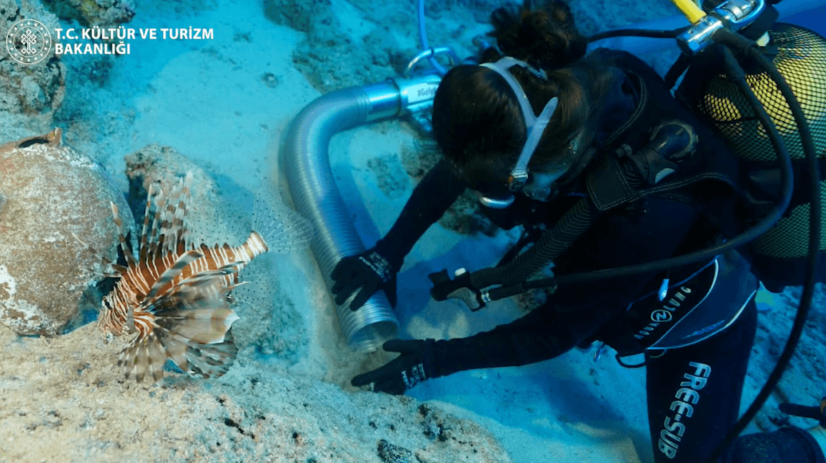
A shipwreck off Türkiye, 4,400 to 4,700 years old, was found with its cargo largely intact. It dates to about when the Great Pyramid was built.
1/5Science News· Brendan Pierson· Oct 14
Tiny implants send signals to each other through body tissue
Georgia Tech engineers built a network that sends small electrical currents through tissue instead of radio. In rats, it linked motion sensors to nerve implants. Implants that only listen use almost no power, so they can be small enough to inject.
Why it matters · Power and size limit what implants can do. A link that costs almost nothing to listen on could let a sensor and a treatment device work together automatically.
Know this first
Context from the editor, not the article · 28 sec
1 · How implants talk today
Connected medical devices, such as an insulin pump paired with a glucose monitor, usually use Bluetooth radio. A radio receiver uses power just by listening, and the body's watery tissue weakens radio waves.
2 · Tissue conducts electricity
Body tissue is full of salty, ion-rich fluid, so it carries small electrical currents. Nerves and the heart run on electrical signals, which is why any current added to the body has to be shown to be safe.
3 · Closed-loop therapy
Doctors have long wanted systems that sense a problem and respond on their own. One example is giving a drug or stimulating a nerve the moment a symptom appears, instead of on a fixed schedule.
2/5Live Science· Kenna Hughes-Castleberry · Oct 17
Mouse study finds the front and back of the brain come from separate cell lineages
A Stanford team tracked mouse embryos and found that the forebrain and the hindbrain grow from two separate groups of early cells. Using that, they grew human hindbrain motor neurons, which labs had long struggled to make.
Why it matters · If it holds up, it changes a textbook picture of how the brain forms and explains a long-running failure in stem-cell labs that has slowed brainstem disease research.
Know this first
Context from the editor, not the article · 30 sec
1 · The brain's three regions
The vertebrate brain has a forebrain (thought, language, planning), a midbrain (relaying senses and movement) and a hindbrain (lower brainstem, which runs breathing, heart rate, swallowing and facial muscles). The brainstem joins the brain to the spinal cord.
2 · Gastrulation and cell fate
Early in development, the embryo goes through gastrulation, when a simple sheet of cells reorganizes into layers that later form organs. Which genes a cell switches on at that point limits what tissue it, and its descendants, can become.
3 · Growing neurons from stem cells
3/5Universe Today· Matthew Williams· Oct 17
Human genome holds its shape after a day of simulated weightlessness
NYU physicists rotated living human cells for 24 hours to imitate weightlessness. The nuclei swelled, but the genome kept its layout and movement, with no DNA damage. Fluid flows from the rotation did damage DNA.
Why it matters · It shows how one hidden flaw in Earth-based microgravity experiments can be removed. It also gives an early, limited answer to whether weightlessness itself changes how our DNA is arranged.
Know this first
Context from the editor, not the article · 34 sec
1 · Packing two meters of DNA
Each human cell nucleus is about 10 micrometers wide but holds roughly two meters of DNA, folded with proteins into chromatin. How it folds affects which genes get used, and folding errors are linked to cancer and developmental disorders.
2 · Faking weightlessness on Earth
Gravity can't be switched off in a lab. A random positioning machine turns samples continuously along two axes, so gravity's pull keeps changing direction and averages out to almost nothing over time. The catch is that the turning sloshes the liquid around the cells.
3 · What spaceflight is known to do
4/5Scientific American· Oct 17
Plant roots bend away from rotting plant tissue, a study finds
Researchers report in Science that roots of Arabidopsis, tomato and wheat grow away from decaying plant matter but not from rotting animal tissue. A hormone that builds up on the side facing the rot drives the turn.
Why it matters · It adds a new sense to the list of things roots respond to, and it uses a different hormone pathway than the textbook model of how plants bend.
Know this first
Context from the editor, not the article · 29 sec
1 · What tropisms are
Plants can't walk away from things, but they can grow in a chosen direction. Tropisms are directional growth responses to cues such as light, gravity, water or salt, made by growing faster on one side of a root or shoot.
2 · How plants usually bend
The textbook mechanism uses the hormone auxin. In light-seeking shoots and gravity-sensing roots, auxin is moved to one side of the organ, cells there grow at a different rate from the other side, and the organ curves.
3 · Abscisic acid's usual job
Abscisic acid (ABA) is best known as a stress hormone. It helps plants cope with drought, for example by closing leaf pores and slowing growth. It has also been linked to how roots grow toward water.
5/5Universe Today· Evan Gough· Oct 14
Remnant of the 1181 supernova shows knotted filaments and an unmoved central star
Astronomers imaged Pa 30, the probable remnant of a supernova seen in 1181. Its gas filaments turn out to be chains of knots, and the star the blast left behind sits almost exactly at the center.
Why it matters · Pa 30 is the only nearby example of a rare, poorly understood kind of stellar explosion, so its shape and its surviving star directly test how these explosions work.
Know this first
Context from the editor, not the article · 32 sec
1 · Two ways stars explode
Core-collapse supernovae come from massive stars dying. Type Ia supernovae come from white dwarfs, dense stellar cores about Earth's size, that blow up completely. Type Iax are a fainter, weaker relative of Type Ia that can leave part of the star behind.
2 · The guest star of 1181
Chinese and Japanese observers recorded a new 'guest star' in the constellation Cassiopeia that stayed visible for about six months in 1181. The nebula Pa 30, found in that part of the sky, is now widely accepted as what the explosion left behind.
3 · Seeing with one element
Hot gas glows at specific wavelengths, called emission lines, that identify the element and its charge. Narrowband filters pass only one line. That lets astronomers map where a single element, such as sulfur or oxygen, sits in a nebula.
The needle-like implant is small enough to inject through a syringe because it doesn't need a large battery.
Engineers at Georgia Tech have shown that tiny medical implants can send signals to each other through the body's own tissue instead of by radio. They call the network 1, short for Smart Wireless Autonomous Networking System. The team reported it on September 24 in Science. So far it has been tested only in rats.
Why radio is a poor fit inside the body
Devices that work together today, such as an insulin pump paired with a glucose monitor, usually use 2, a short-range radio standard. This causes two problems. A radio receiver uses power even while it is only listening, so the device needs a battery, and the battery largely sets how small the device can be. Radio signals also weaken as they pass through tissue, which limits how far apart two devices can be.
Using tissue as the wire
In SWANS, a sender produces an electric pulse. The sender can be a wearable hub on the skin or another implant. The pulse drives a small current through the surrounding tissue, which conducts electricity because it is full of salty fluid. Receiving implants detect that current while using almost no power when they are only listening. Without a large battery, an implant can be small enough to inject through a syringe.
The rat test
The researchers attached motion sensors to rats' front legs and placed 3 on the 4, the main nerve of the back leg. A neurostimulator is a device that delivers electrical pulses to a nerve. When a rat moved a front leg, the external hub sent a signal through its body to the nerve implants, and the back-leg muscles contracted.
Biomedical engineer Alex Abramson says the coordinated legs weren't the point. The test showed that one network can link different kinds of devices: "a 5 showing that you can really connect this system to anything."
The news report gives no figures on power use, data rate, signal range or the number of animals tested.
What comes next
Before any use in people, the team must show that the current doesn't cause harm, such as a disrupted heart rhythm. Abramson says no such problems have appeared in rats so far, but longer tests in larger animals are needed.
The goal is implants that work as a team. One would detect a symptom and another would respond, for example by releasing a drug or stimulating neurons in Parkinson's disease. An external hub, possibly running AI, could coordinate larger networks. Amir Alavi of the University of Pittsburgh, who develops battery-free implantable sensors, says these technologies are now "converging" and could make implants smaller, longer-lived and more connected.
Engineers at Georgia Tech have shown that tiny implants can communicate by sending electrical signals through the body's own tissue instead of by radio. They call the system 1, short for Smart Wireless Autonomous Networking System. The team reported it on September 24 in Science. The tests so far are in rats only, and the source article is a short news piece, so many technical details are missing.
The problem: radio costs power and gets absorbed
Implants are getting smaller and smarter, but getting them to work together is still hard. Devices that coordinate today, such as an insulin pump linked to a glucose monitor, generally use 2, a short-range radio standard. Radio has two drawbacks inside the body.
The first is power. A radio receiver uses energy even when it is only listening for messages. That means a battery, and the battery is often what sets a device's minimum size.
The second is that radio signals weaken as they pass through tissue. That limits how far apart two devices can be and still hear each other.
If a sensor is meant to detect a symptom and tell a second device to treat it, both problems get in the way.
How SWANS works
SWANS uses the body as the wire. A sender, which can be a wearable hub on the skin or an implant, produces an electric pulse. The pulse drives a small current through the surrounding tissue, which conducts electricity because it is full of salty fluid. A receiving implant elsewhere in the body picks up that current.
The important part, according to the article, is that a SWANS implant uses almost no power while it is only listening. Without the need for a large battery, an implant can be small enough to inject through a syringe. The photo shows a metallic, needle-like device.
As a rough analogy (ours, not the researchers'): radio is like shouting across a crowded room, where you have to be loud and every listener has to strain to hear. Sending current through tissue is more like tapping on a shared table. The vibration travels through the material that already connects everyone.
The researchers picture two levels of coordination. Implants could pass simple signals straight to each other. An external hub could manage more complex networks of sensors and treatment devices, possibly using artificial intelligence to decide when to act.
The rat experiment
To test the network, the team attached motion sensors to the front legs of rats. They also implanted 3, devices that deliver electrical pulses to a nerve, on the 4, the large nerve that runs down the back leg. When a rat moved a front leg, the sensor's signal reached the external hub. The hub then sent a signal through the rat's body to the nerve implants, and the back-leg muscles contracted.
Abramson says the coordinated legs were not the goal. The experiment was meant to show that signals could travel through the body and connect different kinds of devices: a motion sensor, a hub and a nerve stimulator. He calls it "a 5 showing that you can really connect this system to anything."
What's missing from the report
The article gives no numbers on how much power the implants use, how fast data moves, how far signals carry, how many animals were tested or how long the tests ran. Those figures decide whether the approach beats radio in practice, so readers who need them should go to the Science paper.
Safety and next steps
Adding electrical current to the body raises an obvious concern. The heart and nerves run on electrical signals, and stray current could interfere with them. Before any use in people, the researchers need to show the signals don't cause problems such as disrupted heart rhythms. Abramson says no such problems have shown up in the rat experiments. But the technology still has to be tested over longer periods and in larger animals, whose bodies differ from rats' in size and in how current spreads.
Where it could lead
Abramson's long-term goal is networks of implants that act on their own. For complex conditions such as Parkinson's disease, an implant could deliver a drug or stimulate neurons at exactly the moment it is needed, triggered by another device that detects the symptom.
Amir Alavi, a materials engineer at the University of Pittsburgh's Swanson School of Engineering, argues the idea gets stronger as related technologies mature: wearables, AI, and miniature battery-free implantable sensors like the ones his own lab develops. "There are now several really interesting directions converging," he says. If they come together, he adds, implants could become "much smaller, simpler, longer-lived, and more connected than what we have today." That is a forecast, not a result. What the work shows so far is that the communication link works in a small animal.
Remember
The main gain is listening power: receivers that use almost nothing while idle don't need large batteries.
The rat demo linked a motion sensor, an external hub and a nerve stimulator into one working loop.
Heart-rhythm safety and long-term tests in larger animals still have to be done before any human use.
Researchers turn human stem cells into specific neuron types by feeding them chemical signals that copy those of the embryo. Forebrain and spinal neurons are fairly routine to make this way. Hindbrain neurons have been notoriously difficult.
Mouse embryo at 9.5 days: the Otx2 lineage (blue) forms the front brain; the Gbx2 lineage (red) forms the hindbrain and spinal cord.
The textbook picture has the brain growing from one continuous sheet of embryonic tissue that gradually divides into front and back regions. A study published in September in 1, a peer-reviewed journal, argues otherwise. Its authors say the brain comes from two separate populations of cells that end up pressed together and look like one organ.
"The front part of the brain and the back part of the brain come from different sources," said Kyle Loh, a developmental biologist at Stanford University and co-author of the study.
Two lineages, two genes
The team studied mouse embryos at 2, the stage when cells first organize into the layers that later form organs. They found two distinct groups of immature cells headed for nervous tissue:
One group switches on 3, a gene that steers early forebrain and midbrain development. It forms the forebrain and part of the midbrain.
The other switches on 4, a gene that steers midbrain and hindbrain development. It forms the hindbrain, the rest of the midbrain, and extends into the spinal cord.
Both genes' roles were already known. What's new is the finding that they mark two separate lineages, which stayed apart instead of mixing as they matured. The two groups also packaged their DNA differently. 5, the protein-and-DNA structure that controls which genes a cell can reach, was arranged differently in each. That points to separate developmental tracks from early on.
Why labs couldn't grow hindbrain neurons
Hindbrain neurons have been hard to make from stem cells. Graduate student Rayyan Jokhai suggests the reason: earlier attempts probably tried to push forebrain- and midbrain-type 6 into becoming hindbrain cells, and the study suggests that can't work. Starting from the correct lineage instead, the team grew functional human hindbrain motor neurons. These cells made proteins typical of the regions that control facial and swallowing muscles.
These are among the neurons that die in 7 (amyotrophic lateral sclerosis) and spinal muscular atrophy, where patients can lose the ability to speak, swallow and breathe. Living neurons can't be sampled from a patient's brainstem, so neurons grown in a dish give researchers a way to watch the diseases happen.
An ancient split, and a speculative story
Using previously published data, the team found the same two-population pattern in macaques, chickens and zebrafish. It also appears in 8, marine invertebrates whose lineage split from vertebrates about 550 million years ago. Loh speculates that the two lineages may once have been two separate nervous systems that merged during evolution. He stresses that "this is only an idea." The study doesn't show when the split arose or how the two parts came together.
The developmental evidence comes mainly from mice. The cross-species comparison reuses existing data, and the article gives no figures on how well the lab-grown neurons work.
A study published in September in 1, a peer-reviewed journal, challenges a basic assumption about how the brain is built. The usual model has the brain arising from a single source: one continuous sheet of embryonic neural tissue that becomes forebrain, midbrain and hindbrain as it grows. The Stanford-led team instead reports that the front and back of the brain come from two distinct populations of cells, which end up side by side.
"The brain appears as one organ," said co-author Kyle Loh, an associate professor of developmental biology at Stanford University. "However, we discovered that the front part of the brain and the back part of the brain come from different sources, which are pushed together to create the appearance of a single organ."
The problem: a part of the brain labs couldn't make
The three regions do different jobs. The forebrain, which includes the cerebral hemispheres, handles language, planning and abstract thought. The midbrain, the top of the brainstem, relays sensory information and helps control movement. The hindbrain, which includes the lower brainstem, coordinates movement and runs breathing, sleep and heart rate.
The hindbrain has been hard to study partly because scientists have struggled to make working hindbrain neurons in the lab. Stem-cell researchers normally build a neuron type by copying the chemical signals an embryo uses. For the hindbrain, that approach has largely failed. The team set out to find out why by going back to the earliest stage at which brain-bound cells can be told apart.
What they found in mouse embryos
The researchers focused on 2 in mice. This is the stage when the embryo's cells reorganize into layers and begin committing to future organs. Among the immature cells destined for nervous tissue, they found two separate populations, each defined by a gene it expresses:
Otx2-expressing cells.3 helps direct early forebrain and midbrain development. In a 9.5-day-old mouse embryo, this lineage forms the forebrain and part of the midbrain.
Gbx2-expressing cells.4 is involved in midbrain and hindbrain development. This lineage forms the hindbrain, the rest of the midbrain, and continues into the spinal cord.
Developmental biologists already knew both genes, including that they label the front and back of the developing brain. The new claim concerns lineage. These are two separate populations that keep their identities as they mature into full nervous tissue and don't overlap. An imperfect analogy: picture a country map drawn as two provinces of one territory, when in fact it was two settlements founded separately that grew until their borders touched.
The team found a second, independent line of evidence in chromatin, the packaging of DNA and proteins inside the nucleus. How tightly a stretch of DNA is packed affects whether a cell can use the genes in it. The two populations had different chromatin configurations. That fits the idea that they follow separate developmental paths, not one shared path that branches late.
Why this explains the lab failures
If hindbrain cells come from their own lineage, the old recipes were probably starting from the wrong material. "Previous attempts to make hindbrain neurons likely tried to coax forebrain and midbrain 5 into hindbrain cells, which our study shows is not possible," said co-author Rayyan Jokhai, a graduate student in Loh's lab. A progenitor is an immature cell committed to producing a certain family of cell types.
Using what they learned, the team guided human stem cells into functional hindbrain motor neurons, the cells that send movement commands to muscles. The neurons produced proteins characteristic of the hard-to-grow hindbrain regions that control facial muscles and the muscles used in swallowing.
This matters for disease research. In spinal muscular atrophy (SMA) and amyotrophic lateral sclerosis (6), motor neurons degenerate, and patients can lose the ability to swallow, speak and breathe on their own. "We cannot sample living motor neurons from a patient's hindbrain to watch how these diseases attack these neurons," Loh said. "Growing these hindbrain motor neurons in a dish gives us a way to investigate these diseases." The team plans to use the cells to study these diseases and look for treatments. The article gives no results from that work yet, and no measures of how closely the lab-grown neurons match real ones.
An old arrangement across species
The researchers also asked whether the split is unique to mammals. Reanalyzing previously published data, they found the same two Otx2 and Gbx2 populations in macaques, chickens and zebrafish. They also found them in 7, marine invertebrates whose evolutionary line separated from vertebrates about 550 million years ago. So the arrangement appears to predate vertebrates.
Anna Di Cosmo, a biologist at the University of Naples Federico II who was not involved, said the work "establishes that the deep ancestry of the vertebrate brain is fundamentally modular." In her view, the separation between the front lineage (forebrain and midbrain) and the back lineage (hindbrain) is "traceably ancient."
The bigger idea, and its limits
Loh's team goes further and speculates that the two lineages were once two separate nervous systems. "Later in evolution, these two nervous systems may have been pushed together to give rise to the appearance of one integrated brain," Loh said. He is careful about it: "It is important to emphasize that this is only an idea, and it might change as scientists learn more."
The evidence supports different claims to different degrees:
Two developmental lineages in mice. This is the core result, supported by gene expression and chromatin data.
Practical payoff. Human hindbrain motor neurons grown from stem cells are reported, but the article gives no numbers on yield or how well they function.
Conservation across species. This rests on existing datasets from other labs, not new experiments.
Two ancestral nervous systems. This is a hypothesis. The study doesn't show when the two pathways arose or how they came to work as one brain.
Several questions remain open. The cross-species pattern should be confirmed with direct lineage tracing, and the lab-grown neurons need to be shown to model ALS and SMA faithfully. Explaining how two separately built tissues wire together at the midbrain to work as one brain is a much harder problem, and the study doesn't address it.
Remember
Otx2 and Gbx2 were already known genes; the new claim is that they mark two separate cell lineages.
Labs probably failed to grow hindbrain neurons because they started from forebrain/midbrain progenitors, which cannot become hindbrain cells.
The idea of two ancestral nervous systems is explicitly speculative; the developmental evidence comes mainly from mice.
Long stays on the International Space Station weaken muscle and bone and affect vision, the heart and the nervous system. NASA's Twins Study compared Scott Kelly, who spent a year in orbit, with his twin Mark on Earth and found changes at the molecular and genetic level.
Human cells, chromatin in green and microtubules in magenta, stretched out after 24 hours of fluid flow.1 / 2
Physicists at New York University held living human cells in simulated weightlessness for 24 hours. The genome stayed almost exactly as it was. The cell nucleus did swell. The only DNA damage came from fluid currents created by the simulation, not from the missing gravity. The study is peer-reviewed and appeared in 1.
The source's headline mentions "long stays in space," but that goes further than the work. This was a one-day experiment on cells in a lab dish on Earth.
The question
The human genome is about two meters of DNA folded into a nucleus about 10 micrometers wide. How it folds affects how genes work, and folding errors are linked to cancer. Gravity presses on all of it constantly. Team leader Alexandra Zidovska wanted to know whether that pressure matters, both here on Earth and for astronauts on long missions.
Faking weightlessness, without the side effects
The team built a 2, which rotates dishes of cells on two axes. "Down" keeps changing direction, so over time gravity's pull on the cells averages out to almost nothing. The weak point of this method is that turning a dish stirs the liquid. The moving fluid pushes on the cells and makes them clump, and according to the article that clumping muddied earlier studies.
The researchers wrote 3D algorithms that steer the machine's motion to keep flow to a minimum. That let them test microgravity and flow separately. After 24 hours under each condition, they recorded microscope images of each cell and its nucleus and looked at the 3 (the membrane around the nucleus), how the genome was arranged and moved, and any DNA damage.
What they found
Simulated microgravity increased the nucleus's volume, which suggests gravity normally compresses it a little.
Flow stretched the cells into longer shapes.
The nuclear envelope's thickness and structure didn't change.
The genome's organization and motion didn't change.
Microgravity caused no DNA damage. Flow did.
The article reports no effect sizes or cell types.
"The genome, its organization, and dynamics are incredibly robust," Zidovska says, at least over 24 hours.
Caveats
A day is short compared with a six-month space station tour or a trip to Mars, and the authors say the small changes they saw could grow over time. An RPM averages gravity away rather than removing it. The test used isolated cells, not whole bodies, and did not include space radiation, which Zidovska notes could still cause damage. The clearest lesson may be about method: earlier RPM studies that found DNA damage may have been partly measuring the stirring of the liquid.
A team at New York University's Center for Soft Matter Research has tested whether gravity shapes how the human genome is physically arranged inside a cell. They simulated weightlessness on Earth for 24 hours. The genome hardly changed. The cell nucleus did grow larger. And the one source of DNA damage was not weightlessness. It was the fluid currents that the simulation creates. The paper appeared in the peer-reviewed journal 1.
The Universe Today headline talks about "long stays in space," which overstates things. This was a one-day experiment on cells in a dish, on the ground, using simulated microgravity. It was not a study of astronauts.
The question
The Human Genome Project finished reading the roughly 3 billion base pairs of human DNA in 2003. A sequence does not tell you how the molecule is arranged in space, though. About two meters of DNA is folded into a nucleus around 10 micrometers wide, and that folding is tied to function. Distorted folding is linked to cancer and developmental disorders. The physical rules behind the folding are still poorly understood.
On Earth, gravity is one constant mechanical force acting on all of this. "To uncover [its role], you have to remove gravity as a force," says Alexandra Zidovska, the NYU physicist who led the work. The answer matters in two places. It matters on Earth, where gravity is always present, and on long missions to the Moon or Mars, where astronauts will spend months or years without it. Space station research and NASA's 2 have already shown that spaceflight affects the body down to the genetic level, but no one yet knows how far those effects go.
Why it's hard to fake zero gravity
Real microgravity requires going to orbit, so most experiments use a ground-based stand-in. The team built a 3. It holds dishes of living cells and rotates them on two axes along a three-dimensional path. Gravity still pulls on the cells, but its direction relative to them keeps changing. Averaged over time, the cells feel almost no net pull in any one direction.
An analogy, which is ours rather than the article's: picture a marble in a bowl that someone keeps tilting in random directions. The marble never settles anywhere, because "down" keeps moving.
This method has a known flaw. Rotating a dish of liquid stirs it. The fluid flows push on cells with a force that real orbit does not produce. The flows also encourage cells to clump into aggregates. The article says these clumps blurred the microgravity signal in earlier studies. That creates a problem of interpretation: any change you see could come from missing gravity or from the stirring.
What the NYU team did differently
The researchers wrote 3D algorithms to drive the machine's motion in ways that keep fluid flow to a minimum. They also wrote algorithms to study what the flows do to cells. That let them run two separate conditions, simulated microgravity with low flow and deliberate exposure to flow. Those conditions could then be compared. According to the article, the approach cut both the flows and the clumping.
After 24 hours in either condition, they imaged the cells with advanced microscopy and recorded image sequences. They measured:
the shape of each cell and its nucleus
the thickness and structure of the 4, the double membrane around the nucleus
how the genome was organized inside the nucleus
how the genome moved over time
signs of DNA damage
One published image shows cells with 5 labeled green and 6, the cell's internal scaffolding, labeled magenta. After 24 hours of flow the cells are visibly stretched out.
Results
The nucleus got bigger. Simulated microgravity increased nuclear volume. The team reads this as a sign that gravity normally squeezes the nucleus slightly.
Flow stretched the cells. Cells exposed to fluid flow became elongated.
The nuclear envelope didn't change. Its thickness and structure were the same in all conditions, which suggests gravity has little effect on it.
The genome held steady. The organization and motion of the genome inside the nucleus were unchanged after 24 hours.
Flow damaged DNA. Microgravity didn't. Simulated weightlessness caused no DNA damage. Exposure to flow did.
The article gives no effect sizes, cell types or sample counts, such as how much the nucleus grew. Readers who want the magnitudes will need the paper itself.
"Our data show that the genome, its organization, and dynamics are incredibly robust and seem unaffected by gravity, or lack thereof, after 24 hours," Zidovska says.
What it means and what it doesn't
The finding about method may be the most useful one. If fluid flow, and not weightlessness, damages DNA in rotating-machine experiments, then earlier RPM studies that reported DNA damage may partly have been measuring the stirring. The article doesn't name specific earlier studies, so it isn't clear which results would be affected.
The finding about biology is reassuring but narrow. Over one day, the physical structure of the genome resists the loss of gravity. Several limits apply:
Time. Twenty-four hours is short next to six-month space station tours or a trip to Mars of several years. The authors say the subtle changes they saw "could amplify over time."
Simulation. An RPM averages gravity away. It does not remove it. Some biologists question how well the method reproduces real orbit.
Radiation. Space has other hazards. Zidovska says structural changes could still happen "due to DNA damage occurring in space," and cosmic radiation is the main known source of that damage. This experiment did not include radiation.
Cells, not people. Isolated cells in a dish lack the tissues, blood flow and hormones of a body.
The team's next steps aren't described in the article. Obvious follow-ups would be longer runs, tests that combine microgravity with radiation, and comparisons with cells flown on the space station. In Zidovska's framing, this study is a first step: it isolates a single force on a single timescale, and longer missions will need answers well beyond it.
Remember
The fluid stirring in Earth-based weightlessness simulations, not the missing gravity, caused the DNA damage seen here.
Gravity appears to compress the cell nucleus slightly, because nuclei grew larger without it.
One day of cells in a dish says little about months in orbit with cosmic radiation.
Plants can't walk, but their roots and shoots bend toward or away from light, water, salt and gravity. These directional growth responses are called 1. A team led by Yuzhou Zhang, a biologist at Northwest A&F University in China, now reports a new one in the journal Science: roots bend to avoid dead or rotting plant tissue. They call it 2.
A spoiled plate
The finding started with a ruined experiment. Zhang was studying how roots respond to gravity in a lab in Austria when airborne microbes contaminated one of his 3, the gel-filled dishes used to grow seedlings. Just before throwing it out, he noticed that almost every root on the plate had bent away from the spoilage. The consistency, he says, "immediately made me wonder whether this was more than a random response."
Testing it on purpose
The team grew 4, a small weed that is plant biology's standard lab organism, in containers. Some containers had patches of rotting apple in them. Roots that met these "decay zones" turned away. Roots in containers with no decay grew straight. Tomato and wheat roots behaved the same way. Rotting animal tissue, though, had no effect.
The researchers suggest the response helps roots avoid disease-causing microbes, much as a bad smell keeps animals away from spoiled food. That is their interpretation of the behaviour. The article doesn't say it was tested directly.
A different hormone
Most familiar tropisms run on the hormone 5. To respond to gravity or light, a plant moves auxin to one side of a root or stem. That changes how fast each side grows, and the organ curves. Saprotropism "does not follow the classical mechanism," Zhang says.
Decaying plant matter makes the soil around it more acidic, and the chemical gradient it creates is different from the one rotting animal tissue leaves. When a root meets that acidified zone, the hormone 6, best known for helping plants cope with drought, builds up on the side facing the rot. Growth on that side is held back, so the root bends the other way.
What's still unclear
Chris Wolverton, a plant biologist at Ohio Wesleyan University who wasn't involved, calls the study's scope impressive. He also says there is "still a lot of open space in the signaling mechanism." One open question is when decay stops being a hazard and becomes useful compost that roots should move toward. Zhang's team plans to study these gaps next.
Plants look stationary, but they move by growing. Roots and shoots bend toward or away from water, light, salt and gravity, and these directional responses, called 1, are central to how a rooted organism survives. In a paper in the journal Science, a team led by Yuzhou Zhang of Northwest A&F University in China describes a new one. Roots bend away from dead or rotting plant tissue. The researchers call it 2, from the Greek sapros, "rotten."
An accident worth noticing
The discovery began with a failed experiment. Zhang was working in a laboratory in Austria on 3, the way roots grow downward in response to gravity. One of his 4, the gel-filled dishes used to grow seedlings where their roots can be seen, got contaminated with airborne microbes. Contaminated plates normally go straight in the bin. Before throwing it away, Zhang saw that almost every root on the plate had bent away from the spoiled area.
The uniformity was what caught his attention. A few roots wandering off course would mean nothing, but nearly all of them turning the same way pointed to a real response. It "immediately made me wonder whether this was more than a random response," he says.
Turning an observation into an experiment
To check it, the team grew 5, a small mustard-family weed that is the standard model organism of plant biology, in containers. Some containers held patches of rotting apple, which the researchers called "decay zones." The other containers had none and served as controls.
Roots that reached a decay zone bent away from it.
Roots in containers without decay grew straight through.
Tomato and wheat roots showed the same avoidance. These are distant relatives of Arabidopsis, and wheat is a grass, so the response isn't limited to one plant lineage.
Putrefying animal tissue had no effect on root direction.
That last result matters most. It shows the roots weren't simply avoiding anything rotten or microbe-covered. They responded specifically to decaying plant material. The article doesn't give counts, bending angles or statistics, so it isn't possible to say here how strong or how consistent the effect was across species.
Zhang and his colleagues propose that the behaviour helps plants avoid pathogens, since rotting plant tissue is likely to carry microbes that attack plants. They compare it to the way an unpleasant smell steers animals away from spoiled food. The article presents this as the team's theory. It doesn't report experiments showing that saprotropic roots actually suffer less infection.
Why the mechanism is unusual
Most well-studied tropisms run on the hormone 6. In the classical picture, when a root senses gravity or a shoot senses light from one side, auxin is moved to one flank of the organ. Cells on that flank grow at a different rate from cells on the other, and the organ curves. A rough analogy: two rows of bricklayers building a wall side by side, where one row works faster. The wall leans toward the slower side.
Saprotropism "does not follow the classical mechanism," Zhang says. The team found it depends instead on 7, a hormone best known as a stress signal that helps plants cope with drought and other hardships. As described in the article, the sequence runs like this:
Decaying plant matter acidifies the area around it. The chemical gradient it creates differs from the one decaying animal matter produces, which may explain why the roots tell the two apart.
A root growing into that gradient experiences it more strongly on the side facing the rot.
ABA accumulates on that side.
The asymmetry pushes growth in the other direction, and the root curves away.
So the logic is the same as other tropisms: an uneven cue leads to an uneven hormone, which leads to uneven growth. What differs is the hormone doing the work. The article doesn't say how the root senses the acidity, whether auxin plays any supporting part, or which genes are involved.
Reception and open questions
Chris Wolverton, a plant biologist at Ohio Wesleyan University who wasn't involved in the work, says the study's scope is impressive. He also says "there's still a lot of open space in the signaling mechanism for more detail to get fleshed out."
He raises one example. Decaying plant matter isn't only a hazard. Broken down far enough, it becomes compost, a source of nutrients roots would want to reach. At what point does rot stop being something to avoid and become something to grow toward? The study as reported doesn't answer that, and the answer would determine how saprotropism plays out in real soil. There, dead leaves and roots are everywhere and at every stage of decay, unlike a container with a lump of rotting apple.
Other gaps follow from that:
Field relevance. The experiments used containers and, at least at first, agar plates. Whether roots in natural soil, with its patchy chemistry and microbes, show the same avoidance isn't addressed.
The benefit. The pathogen-avoidance idea is plausible but, as far as the article reports, untested.
The trigger. Acidification is named as the cue, but other chemicals released by decay might also play a part.
Zhang's team says it plans to work on these gaps. The work has been peer-reviewed and published in a major journal, but this is the first description of the phenomenon. Independent replication and the missing parts of the signaling pathway are still to come.
Remember
Roots avoided rotting apple but ignored rotting animal tissue, so they respond specifically to plant decay.
The bending runs on abscisic acid, not auxin, the hormone behind the classic light and gravity responses.
It is untested whether avoiding rot protects plants from disease, or when decay turns into useful compost.
Gemini North image of Pa 30 through the sulfur filter, showing filaments made of chains of knots.1 / 3
In 1181, observers in China and Japan recorded a "guest star" in Cassiopeia that shone for six months. Its debris is almost certainly the nebula Pa 30. A peer-reviewed study in The Astrophysical Journal, led by Tim Cunningham of the University of Warwick, gives the most detailed view of it yet.
Why this remnant matters
Pa 30 is thought to come from a 1, a weaker relative of the Type Ia explosions in which a 2 (the dense leftover core of a Sun-like star) blows apart. In a Type Iax event, part of the star can survive. Researchers think this one was triggered by two white dwarfs merging. Pa 30 is the only known remnant of this kind in the Milky Way. It is close, and its age is known exactly, which makes it a natural laboratory for how such explosions spread material into space.
Knots, not streaks
The team used 3 (the Gemini Multi-Object Spectrograph) on the Gemini North telescope to take deep images through 4 filters. These pass only light from one element in one charge state. Light from singly ionized sulfur, written 5, showed far more structure than earlier images taken in doubly ionized oxygen, 6. The authors count about ten times more filaments than before.
The filaments also aren't smooth. They are "cascades of knots or clumps," like beads on a string (our analogy). The team reads this as a sign that the gas around the star was patchy in density and temperature. As the debris slammed into it, the patches broke the outflow into clumps.
A star that wasn't kicked
The surviving white dwarf is extreme. It may weigh about 1.5 solar masses, above the roughly 1.4-solar-mass limit for ordinary white dwarfs. It is about 200,000 °C, and its wind blows at a record 16,000 km/s.
Many Type Iax models predict a lopsided explosion that should kick the survivor sideways, away from the center of the debris. The team estimated the nebula's center in 1181 and in 2025 and found both consistent with the star's position. In other words, there was no large sideways kick. Any model of Pa 30 now has to explain this, along with the knotty filaments.
Caveats
The measurement covers only sideways motion across the sky, not motion toward or away from us. The merger origin is still a hypothesis. The authors say Hubble's sharper resolution is needed to firm up the analysis. Since reliable records of stellar explosions go back only about a thousand years, Cunningham expects similar remnants are still waiting to be found in our galaxy.
A remnant with a written record
In 1181, observers in China and Japan wrote down a "guest star" in Cassiopeia that shone for about six months. They left three independent records of it. Astronomers are now nearly certain they have found the debris: a nebula called Pa 30 (also listed as IRAS 00500+6713). A new peer-reviewed study in The Astrophysical Journal (vol. 1010, p. 28), led by Tim Cunningham of the University of Warwick and the Center for Astrophysics | Harvard & Smithsonian, gives the sharpest look yet at its structure. The team reports two main findings. The ejecta filaments are chains of small knots, not smooth streaks. And the surviving star sits almost exactly at the nebula's center.
Why Pa 30 is special
Most supernovae fall into two families. Core-collapse supernovae happen when massive stars run out of fuel. Type Ia supernovae happen when a 1, the dense, burnt-out core of a Sun-like star, undergoes runaway nuclear burning and is usually destroyed outright. Type Iax is a weaker, fainter subclass of Type Ia. In many models, the burning fails to fully detonate the white dwarf, so part of the star survives.
Researchers think the 1181 event was a 2, triggered when two white dwarfs merged. That makes Pa 30 the only known remnant of this kind in the Milky Way. (The Universe Today article says it is the only known supernova remnant in the galaxy, which is clearly an error. Hundreds are known.) Because Pa 30 is close and its age is known from the historical records, it is a rare chance to watch how such an explosion throws material into space. That material eventually ends up in new stars.
What the new images show
The team used the Gemini Multi-Object Spectrograph (3), an instrument on the 8-metre Gemini North telescope in Hawaiʻi, to take deep 4 images. A narrowband filter passes only a thin slice of wavelengths. Pointed at one emission line, it shows where a single type of glowing gas sits. The key line here was 5, from sulfur atoms that have lost one electron. It is a standard tracer of ionized gas.
Earlier images of Pa 30 mostly used 6, from oxygen that has lost two electrons. When the team lined up [S ii] and [O iii] images of the same areas, the sulfur images showed far more structure. The authors report roughly ten times more filaments than earlier observations found.
They also found a new kind of structure. "The filaments are not smooth radial streaks, but instead are composed of cascades of knots or clumps," the researchers write. One way to picture it (an analogy, not the authors'): the filaments look less like jet contrails and more like beads on a string. The team reads the knots as a record of the surroundings. The ejecta ploughed into the 7, the gas and dust already around the star. Patches of different density and temperature in that gas broke the outflow into clumps as it expanded.
Co-author Ilaria Caiazzo of the Institute of Science and Technology Austria credits Pa 30's closeness to Earth for the level of detail.
The star that stayed put
Pa 30 has something most Type Ia remnants cannot have: a surviving star. Its central white dwarf is extreme. It may be "super-Chandrasekhar," meaning it is heavier than the roughly 1.4-solar-mass limit, called the 8, above which an ordinary white dwarf cannot support itself. Its estimated mass is about 1.5 solar masses. It is also very hot, about 200,000 °C, and it drives a stellar wind at about 16,000 km/s, a record speed.
Its position is also unusual. In many Type Iax models, the explosion is lopsided: a failed detonation, or a partial deflagration (subsonic burning) that starts off-center. "The surviving remnant can receive a substantial recoil kick," the authors note. A star that was kicked sideways in 1181 should have drifted away from the point the debris is expanding from.
To test this, the team estimated where the nebula's center was in 1181 and where it is in 2025. They then compared those positions with the central star, using uncertainty contours at 1, 2 and 3 sigma. Sigma is the standard measure of statistical confidence, and 3 sigma is the strictest of the three. The result: "We measure a nebula center consistent with the position of the central star, indicating that the remnant has not received a large transverse kick." "Transverse" means sideways across our line of sight. The article does not give the measured upper limit on the kick speed.
Why it matters, and what's missing
Both findings narrow down which explosion models can work. A model for Pa 30 now has to produce knotty filaments, and it has to leave a heavy, very hot survivor without a strong sideways kick. That favors explosions that were fairly symmetric, or models where the kick is small. The authors say the shape and the recoil together constrain the progenitor, the system that exploded. But they also say firm conclusions need better data. "Further observations exploiting the exquisite angular resolution of the Hubble Space Telescope would greatly enhance this analysis," they write.
Some caveats apply. The kick measurement only covers sideways motion, so it says nothing about motion toward or away from us. The link between the knots and the circumstellar medium is an interpretation of the images, not a direct measurement. The white dwarf merger origin is the leading hypothesis but has not been proven. The article also does not explain how the 1181 nebula center was reconstructed. Presumably the team ran the filaments' motion backward, but that is not stated.
More out there?
Cunningham points out that reliable records of stellar explosions go back only about a thousand years, a tiny fraction of the galaxy's history. "There might well be similar remnants still lurking in the dark in our own galaxy," he said, and he expects new telescopes to find them.
Remember
Images in sulfur light reveal about ten times more filaments in Pa 30 than earlier oxygen-light images did.
The filaments are chains of knots, which suggests the debris hit patchy surrounding gas.
The surviving white dwarf sits at the nebula's center, which rules out a large sideways recoil from the explosion.